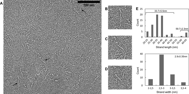
FIGURE 2.
Electron microscopy of fragment 760–1733. A–D, negative staining observation. The scale bar for A has a length of 100 nm. Several different morphologies are observed. Objects indicated by a black arrow are enlarged in B–D. E, histograms of the measured lengths (top) and widths (bottom) of the fibers.

