
FIGURE 7.
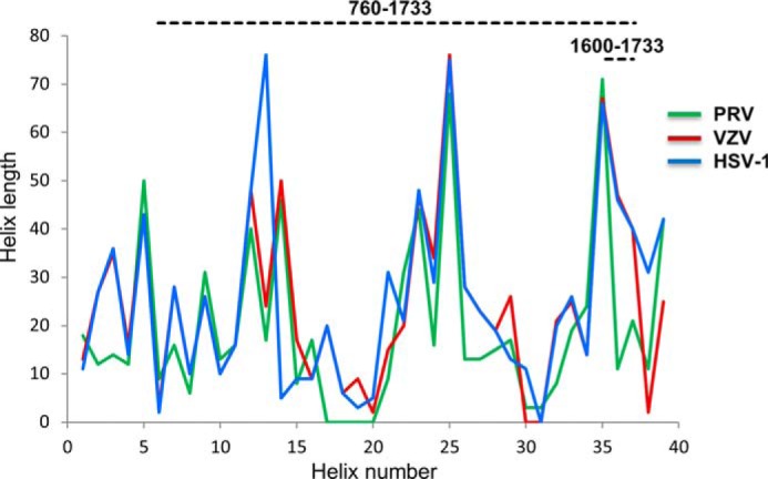
Secondary structure of the central stalk is strongly constrained across alphaherpesvirinae. The secondary structures of fragments 584–1836 of HSV-1 (blue), 522–1708 of VZV (red), and 398–1419 of PRV (green) UL36 were predicted using SOPMA. Plotted are the predicted lengths of successive helices (numbered 1–39 from N to C terminus) in these fragments. Three insertions are plotted as helices of length 0 in viruses where they are not present (helices 17–20 in PRV, helix 31 in HSV-1, and helices 30–31 in VZV). The HSV-1 and VZV curves are superimposed between helix numbers 4 and 12.