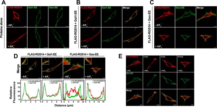
FIGURE 1.
RGS14 is recruited to the plasma membrane by inactive Gαi1-GDP and by active Gαo-AlF4−. A, HeLa cells were transfected with either 100 ng of FLAG-RGS14 or 100 ng of EE-tagged Gα proteins. Transfected cells were treated with AlF4− for 10 min prior to fixation for immunofluorescence and confocal microscopy as described under “Experimental Procedures.” Images are representative of three separate experiments. Scale bar, 10 μm for all panels. B, HeLa cells were co-transfected with 100 ng of FLAG-RGS14 and 100 ng of Gαi1-EE. Cells were treated with AlF4− and fixed as in A. Images are representative of three separate experiments. C, HeLa cells were co-transfected with 100 ng of FLAG-RGS14 and 100 ng of Gαo-EE. Cells were treated with AlF4− and fixed as in A. Images are representative of three separate experiments. D, intensity graphs indicating relative fluorescence from merged images in B and C. Relative fluorescence intensity of FLAG-RGS14 and either Gαi1-EE or Gαo-EE was measured at the plasma membrane as indicated by the white line in merged images. Intensity graphs were generated in ImageJ and are plotted from left to right. Insets highlight the plasma membrane in each image. E, HeLa cells were transfected with 100 ng of FLAG-RGS14 (top row) and 100 ng of EE-tagged Gαo (middle row) and treated with AlF4− for 0, 2, 5, and 10 min prior to fixation for immunofluorescence and confocal microscopy as described under “Experimental Procedures.” Merged overlay images of RGS14 and Gαo are shown (bottom row). Scale bar, 10 μm. Note that images for the zero and 10 min time points are from C.

