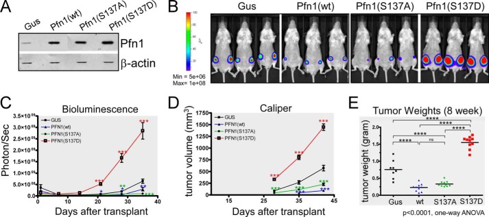
FIGURE 2.
Mimicking Pfn1 phosphorylation on Ser-137 promotes tumor growth in vivo. A, Western blot showing stable overexpression of exogenous Pfn1 (wt, S137A, and S137D) over endogenous Pfn1 in MDA-MB-231 cells. gus, a bacterial reporter gene encoding β-glucuronidase as the negative control. β-Actin was blotted as the loading control. B–E, stable MDA-MB-231 cells were injected into the mammary fat pads of 5-week-old female NOD/SCID mice (106 cells per side, 5 mice per group). Tumor cells were monitored 1 day after injection and weekly thereafter using bioluminescence imaging (BLI). Caliper measurements started 4 weeks after injection. B, representative bioluminescence imaging images showing tumor burden at 4 weeks post-injection. C, time course of tumor growth measured by bioluminescence imaging. Total photon flux (photons/s) of 10 s/binning 2 imaging acquisition is shown for each data point. Data are the mean ± S.E. of 10 ROI (region of interest covering each entire tumor) values for each group (2 tumors per mouse, 5 mice per group). D, time course of tumor volume increase measured by caliper. Data are the mean ± S.E. of 10 tumors within each group (2 tumors per mouse, 5 mice per group). p values were based on two-tailed unpaired t test (Pfn1 versus Gus tumors). E, end-point tumor weights at resection. p values were based on one-way ANOVA and multiple comparisons using the Tukey method. *, p < 0.05; **, p < 0.01; ***, p < 0.001, ****, p < 0.0001.

