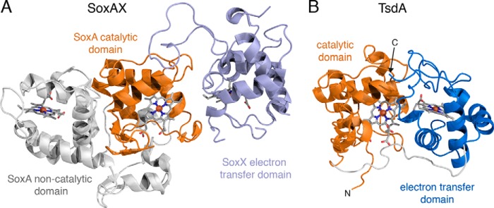
FIGURE 4.

Structural comparison between R. sulfidophilum SoxAX (A) and A. vinosum TsdA (B). The proteins are displayed with their catalytic domains (orange) in the same orientation as determined by an alignment of their Cα atoms. The hemes are shown in stick representation with carbon gray, nitrogen blue, oxygen red, sulfur yellow, and iron orange.
