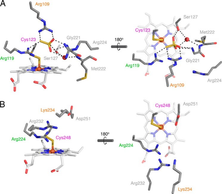
FIGURE 7.
Comparison of the active sites of TsdA and SoxAX. Active sites are viewed from approximately the same orientations as in Fig. 6 (left-hand panels) and Fig. 4 (right-hand panels). Amino acid side chains, hemes, and thiosulfate are shown in stick representations and colored as in Fig. 4. Ordered water molecules are shown as red spheres. Non-covalent bonding interactions are indicated with black dotted lines. Structurally homologous amino acids are labeled in the same color. A, active site of A. vinosum TsdA. Crystals were grown in the presence of tetrathionate, and a thiosulfate molecule is present in the active site. Note that the right-hand heme propionate exhibits an additional minor conformer (not shown) in which the propionate points upward into the active site. B, active site of R. sulfidophilum SoxAX (PDB entry 1H32). Note that for consistency with TsdA and the current PDB numbering convention, the SoxA amino acids are numbered from the start of the precursor protein and thus differ from the numbering scheme used previously (9).

