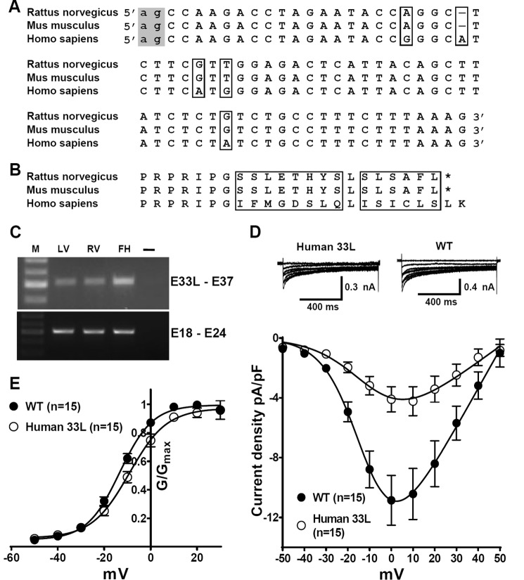
FIGURE 10.
Human Cav1.233L channels conduct Ca2+ ions and are developmentally regulated in heart. A, alignment of the exon 33L DNA sequences from Rattus norvegicus, Mus musculus, and Homo sapiens. The canonical -ag- acceptors (indicated in the gray box) is preserved in all three species. The mouse and the rat share the same exon 33L sequence. Human exon 33L is predicted to contain an additional nucleotides. All the differences in DNA sequences are highlighted by boxes. B, alignment of the amino acid sequences of exon 33L predicted from R. norvegicus, M. musculus, and H. sapiens. The differences in amino acid sequences are labeled by boxes. No stop codon is generated by the inclusion of human exon 33L. C, using human exon 33L-specific primers, a band of 502 bp was found in human adult left ventricle (LV), adult right ventricle (RV), and fetal left ventricle (FH). M, marker. DNA sequencing confirmed the sequence to be human exon 33L. PCR using primers across exon 18–exon 24 was illustrated as the loading control. As a water control (–), cDNA was replaced by water to exclude possible contaminations. D, human exon 33L was cloned into a Cav1.2 reference channel. I-V curves were obtained in a bath solution containing 1.8 mm Ca2+. In HEK cells, human Cav1.233L channels indeed conduct Ca2+ ions with a smaller current density. E, conductance was calculated as G = I/(Vm − ECa) and normalized by the maximum conductance value and fit with Boltzmann equation, where ECa is reverse potential.

