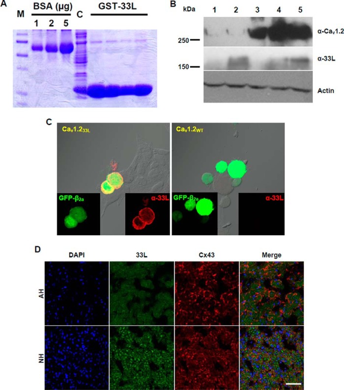
FIGURE 4.
Generation and characterization of a polyclonal antibody against exon 33L. A, The 22-amino acid polypeptides encoded by exon 33L were fused with GST (GST-33L) and purified for rabbit immunization. BSA standards of 1, 2, and 5 μg are shown. M, protein marker. C, crude lysate. B, representative Western blot using the exon 33L-specific antibody (α-33L) showed a band of 160 kDa in both NH and AH. Full-length Cav1.2 channel (∼270 kDa) and actin (42 kDa) were probed as controls. Lane 1, HEK cells transfected with pcDNA3 vector; lane 2, HEK cells transfected with Cav1.233L; lane 3, HEK cells transfected with wild type Cav1.2; lane 4, AH; lane 5, NH. C, immunofluorescent staining of Cav1.233L- or Cav1.2WT-transfected HEK cells with polyclonal antibody α-33L (red). GFP-tagged β2a was co-transfected together with the α1 subunits. D, representative immunofluorescent staining showed exon 33L expression in NH and AH. Connexin 43 (Cx43) was co-stained, and nuclei were labeled with DAPI. Scale bar: 50 μm.

