FIGURE 5.
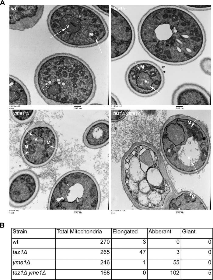
Electron microscopy reveals severe mitochondrial morphological abnormalities in taz1Δ yme1Δ double mutant yeast. Wild type (W) and mutant strains were grown to early stationary phase (0.9–1.0 A600) in SC medium with 2% lactate and then processed for electron microscopy as described under “Materials and Methods.” Briefly, the harvested cells were treated with 1.5% KMnO4 and 1% sodium periodate before they were ethanol-dried and processed for electron microscopy using an LKB Huxley Ultramicrotome. The cells were then viewed using a JEOL JEM 1230 transmission electron microscope, and the images were captured with a Hamamatsu ORCA-HR digital camera. A, panels show a representative EMs. Wild type and taz1Δ yeast harbored predominantly healthy mitochondria with an increase in the presence of healthy but elongated mitochondria in taz1Δ yeast. Both yme1Δ and taz1Δ yme1Δ yeast harbored abnormally shaped mitochondria with vacuolated cristae. These defects were pronounced in taz1Δ yme1Δ cells, which also harbored cells with giant mitochondria, and further, many of the cells were not intact or were dying with no visible mitochondria. Arrows, vacuole (V) and mitochondria (M). B, tabulation of mitochondrial morphologies from at least 150 mitochondria for each strain.

