
Figure 1. PTEN Is Organized along MTs.
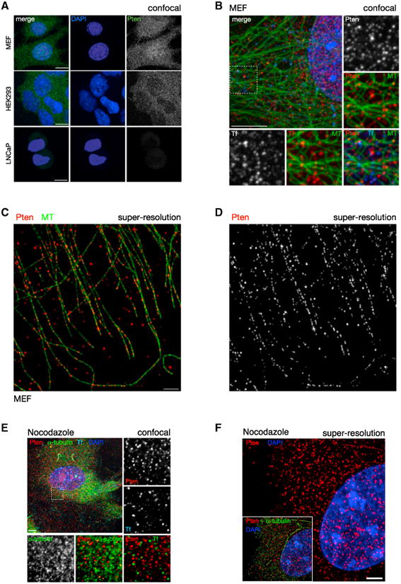
(A) Confocal immunofluorescence (IF) microscopy reveals that PTEN distribution is punctate in the cytoplasm. Punctate distribution is conserved in both primary WT MEFs (top panels) and HEK293 cell lines (middle panels). PTEN null cells (human prostate cancer-derived LNCaP) display minimal background staining, confirming antibody specificity (bottom panels). Images show total projections of all Z sections (extended focus). Scale bars, 10 μm.
(B) Cytoplasmic PTEN distribution is indistinguishable from that of labeled transferrin (Tf). Pten and Tf punctate stains show a similar close association with MTs. Images of WT MEFs, single section (Z = 1). Scale bar, 10 μm.
(C and D) Super-resolution light microscopy (OMX) reveals that PTEN is organized along MTs. Scale bar, 2 μm.
(E) Nocodazole treatment of WT MEFs abrogates MT assembly but does not dissolve the punctae of Pten or Transferrin, compared to alpha-tubulin. Z = 1. Scale bar, 10 μm.
(F) Nocodazole treatment abrogates the linear arrangement of PTEN along MTs. Z = 1. Scale bar, 2 μm.
See also Figure S1.