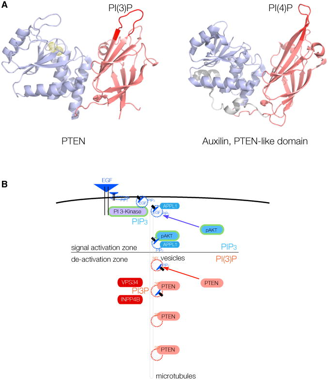
Figure 7. PTEN Vesicle Binding May Be an Evolutionarily Conserved Function in Endocytosis.

(A) Left: phosphatase (blue) and C2 domain (red) of PTEN, with the essential CBR3 PI(3)P-binding loop highlighted (left, crimson). Right: structure of the PTEN-like region of Auxilin, with the essential loop for PI(4)P binding highlighted (right, crimson).
(B) Model for spatio-temporal maturation of vesicle-associated PIP3-lipid signaling through APPL1-positive vesicles (signal activation zone)and subsequent PIP3 signal termination by PTEN after generation of PI(3)P (also labeled 3P) onto vesicle membranes by, e.g., VPS34 or INPP4B (de-activation zone).
