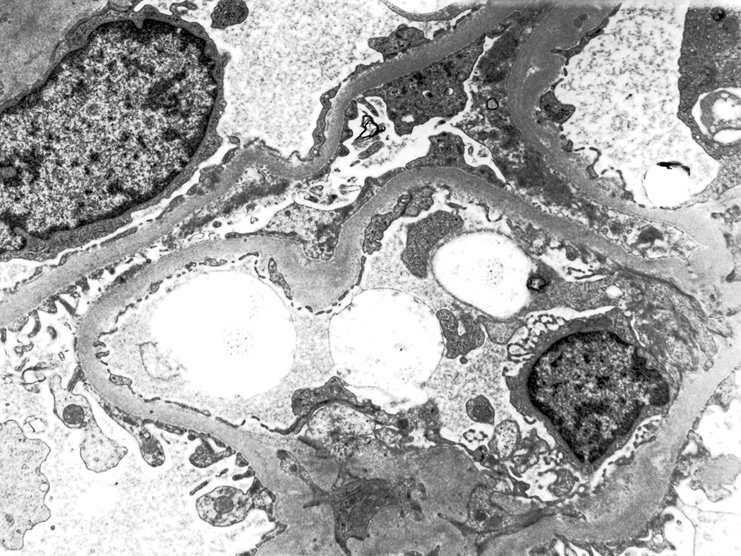
Figure 5.
Electron micrograph of the kidney biopsy of a 42-year-old man with type 2 diabetes mellitus, hemizygous for the GLA p.(Arg118Cys) sequence variant, identified in the PORTYSTROKE study. The patient presented with mild proteinuria and a kidney biopsy was obtained for the differential diagnosis with diabetic nephropathy. Light microscopy examination was diagnostic of diabetic nephropathy and the electron microscopy study did not show any intracellular inclusions with the “zebra body” or “myelin figure” morphology, typical of Fabry disease nephropathy.

