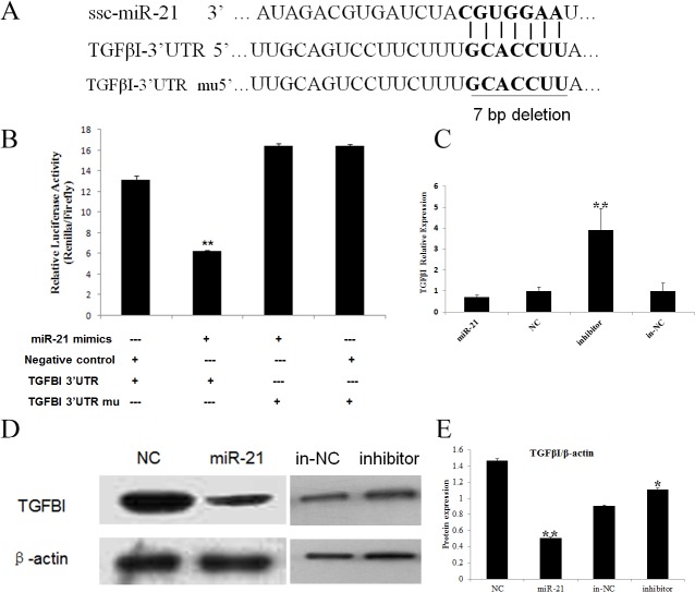
Fig 3. TGFβI is a target for miR-21.
A. The predicted binding site between miR-21 and the TGFBI 3' UTR. B. Detection of luciferase activity in the PIEC cells after co-transfection with the luciferase reporter and miR-21/NC. The PIEC cells were co-transfected with the psi-check2-TGFβI 3’ UTR and miR-21 mimics/negative control duplex. The relative luciferase activity was measured after 48 h. The data represent the mean ± SD from three independent experiments performed in duplicate. C. RT-qPCR was performed to verify the decreased TGFβI expression following the overexpression of miR-21 and enhanced expression after miR-21 inhibition in the PIEC cells. The data represent the mean ± SD from three independent experiments performed in duplicate. D. Total cell extracts were harvested at 48 h after transfection, and the TGFβI protein was analyzed via immunoblotting. E. All quantitative results were normalized byβ-actin and were shown as mean ± SD from three independent experiments.

