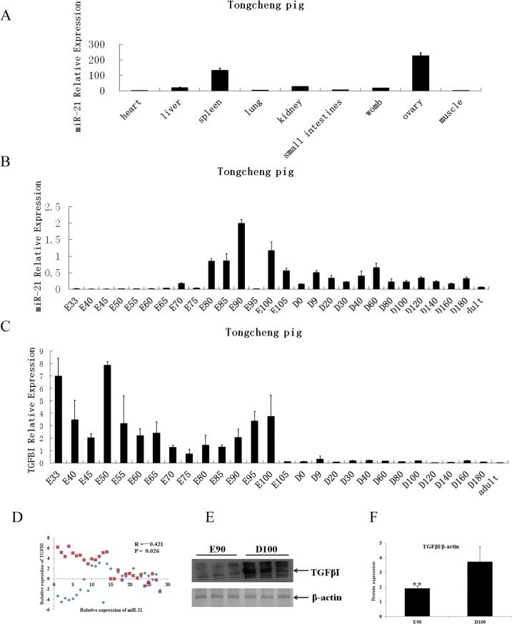
Fig 4. Expression profiles of miR-21 and TGFβI in Tongcheng pigs.
A. Tissue distribution of miR-21. Total RNAs were extracted from the heart, liver, spleen, lung, kidney, small intestine, uterus, ovary and longissimus dorsi tissues. B. Expression profiles of miR-21 at different developmental stages. Total RNAs were extracted from the longissimus dorsi of the Tongcheng pigs at different dpcs (E) and postnatal days (D). The relative expression of miR-21 was compared to the internal control reference U6 and was analyzed using ΔΔCt. The data represent the mean ± SD from four independent experiments performed in duplicate. C. Expression profiles of TGFβI at different developmental stages. Analyses were performed according to B. D. Correlation between miR-21 and TGFβI expression during myogenesis in pigs. E. TGFβI protein levels were analyzed in extracts from the longissimus dorsi on E90 and D100 via immunoblotting. F. All quantitative results were normalized byβ-actin and were shown as mean ± SD from three independent experiments.

