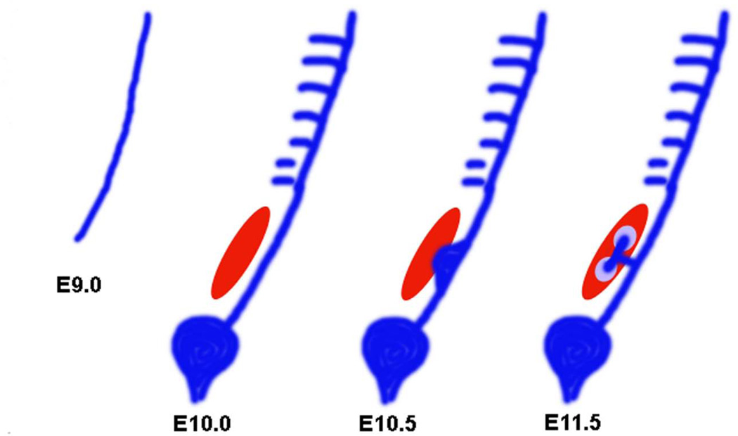
Figure 1. Stages of kidney development in the mouse embryo.
The pronephric duct arises from the intermediate mesoderm in the mouse embryo at E9.0. The nephric duct grows caudally until it reaches the cloaca. Mesonephric tubules can be seen at E10.0. The mesonephric tubules which are found more caudally, are more developed compared to the proximal ones. Posterior cells of the intermediate mesoderm specialize to form an aggregate called metanephric mesenchyme (red). The metanephric mesenchyme gives rise to all the segments of the nephron. The ureteric bud, an outgrowth from the nephric duct invades the MM by E10.5. The UB gives rise to the collecting system of the kidneys. The UB bifurcates and the induced mesenchyme, known as the cap mesenchyme (purple), surrounds the tips of the UB.

