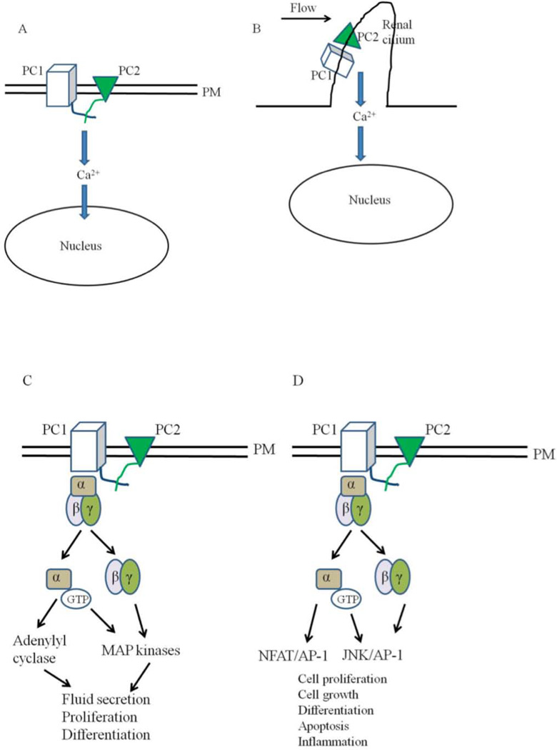
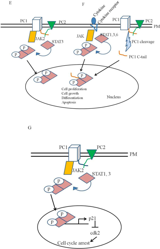
Figure 4. Models for the functions of polycystins.
A: PC1 and PC2 are found on the plasma membrane (PM) in this model. Activation of PC1 leads to the activation of PC2 which leads to calcium influx and changes in gene transcription. B: Polycystins signaling through the primary cilia. Renal cilia bend in response to fluid flow. Bending of the cilia leads to the influx of calcium through polycystins. C and D: Polycystins regulating G-protein signaling. Binding of PC1 to heterotrimeric G proteins activates the Gα subunit and the release of the βγ subunits. This leads to the activation of adenylyl cyclases and MAP kinases which can affect several cellular processes such cell proliferation, fluid secretion and differentiation. G protein signaling through polycystins can also affect the JNK/AP-1 and NFAT/AP-1 pathways. E: Polycystins signaling through the JAK-STAT signaling pathway. E: Membrane anchored PC1binds to JAK2 and activates it which then phosphorylates STAT3. F: PC1 C tail can translocate to the nucleus and coactivate STAT1, 3 and 6 which were already activated independently through cytokine signaling. Once in the nucleus, STAT transcription factors affect the expression of genes required for cell proliferation, cell growth, differentiation and apoptosis. G: Polycystins regulating the JAK-STAT signaling pathway. PC1, in a reaction requiring PC2, activates JAK2 which leads to the phosphorylation of STAT1. Phosphorylated STAT1 translocates to the nucleus and activates the cyclin-dependent kinase inhibitor, p21. Activation of p21 results in the subsequent inhibition of cdk2 and cell cycle arrest.


