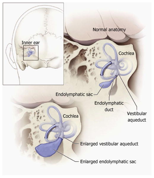
Figure 1. Schematic illustration of the relationship of the vestibular aqueduct with the endolymphatic sac and duct.

Normal anatomy of the inner ear structures is shown above. Pathologic enlargement of the endolymphatic sac and abnormal enlargement of the vestibular aqueduct are shown below. Some ears with enlargement of the vestibular aqueduct also have a reduced number of cochlear turns. Reproduced from http://www.nidcd.nih.gov/health/hearing/vestAque.htm.
