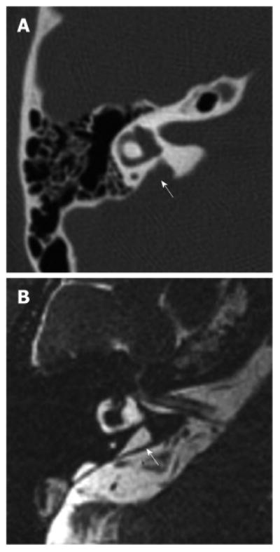
Figure 2. Right temporal bone of a patient with enlargement of the vestibular aqueduct.

A: Axial computer tomography image of a right temporal bone with an enlarged vestibular aqueduct (arrow); B: Equivalent magnetic resonance image of the same temporal bone showing an enlarged endolymphatic duct (arrow). Reproduced from http://www.nidcd.nih.gov/health/hearing/Pages/eva.aspx.
