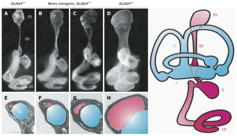
Figure 3. Morphology of the endolymphatic sac and duct and vestibular aqueduct in Slc26a4 mutant mouse models of enlargement of the vestibular aqueduct.

Slc26a4Δ/+ normal control (A and E), binary transgenic; Slc26a4Δ/Δ (B, C, F and G), or Slc26a4Δ/Δ mutant control mice (D and H) were sacrificed at P3 for paint-fill analysis (A-D) or between P28 and P109 for cross-sectional histopathology of the vestibular aqueduct (VA, shaded pink) adjacent to the common crus (CC, shaded blue; E–H). Scale bars: 500 μm (A, applies to A–D; E, applies to E–H). Manipulating pendrin expression in binary transgenic; Slc26a4Δ/Δ mice results in less enlargement of the endolymphatic duct and sac and vestibular aqueduct (B, C, F and G). ES: Endolymphatic sac; ED: Endolymphatic duct; S: Saccule; U: Utricle; CO: Cochlea. Reproduced with modification from Choi et al24.
