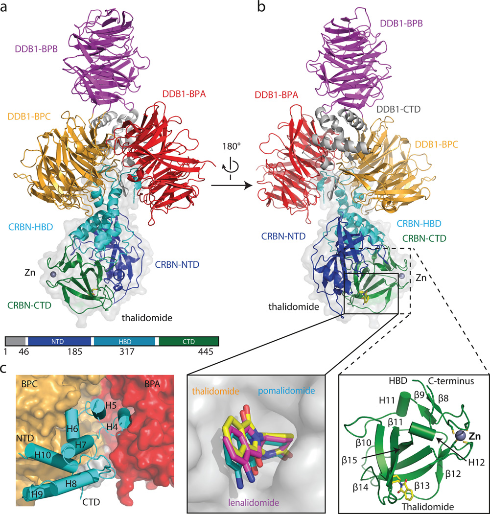
Figure 1. Overall structure of the DDB1-CRBN complex.
(a) Cartoon representation of the hsDDB1-ggCRBN-thaliomide structure: DDB1 highlighting domains BPA (red), BPB (magenta), BPC (orange) and DDB1-CTD (grey); ggCRBN highlighting domains NTD (blue), HBD (cyan) and CTD (green). The Zn2+-ion is drawn as a grey sphere. (b) As in (a) with the thalidomide shown as yellow sticks. A close-up showing that all IMiDs occupy a common binding site on CRBN, and a close-up of the overall ggCRBN-CTD architecture. (c) ggCRBN-HBD helices and their interactions with DDB1.

