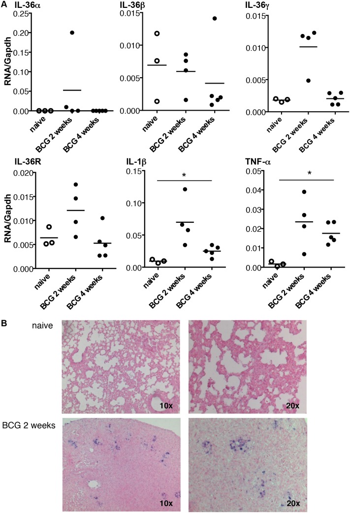
Fig 2. Kinetic of IL-36 cytokines, IL-36R, IL-1β and TNF-α mRNA expression after M. bovis BCG infection.
mRNA levels of Il36a, Il36b, Il36g, Il36r, Il1b, and Tnfa measured by RT-qPCR and normalized to Gapdh mRNA levels from the lungs of C57BL/6 wild-type mice 2 weeks (n = 4) and 4 weeks (n = 5) after M. bovis BCG infection. Three naïve mice were included as controls (A). In situ hybridization was performed using a digoxigenin-labeled riboprobe complementary to Il36g mRNA (B). The micrographs are representative of lungs from one naïve mice (upper panel) and one infected mice 2 weeks after M. bovis BCG inoculation (lower panel). *P<0.05, as assessed by Kruskal-Wallis, followed by Mann-Whitney test.

