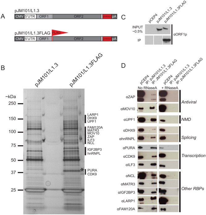
Fig 1. The identification of host proteins immunoprecipitated with L1 ORF1p-FLAG.
(A) Schematic of L1 constructs: pJM101/L1.3 expresses a human L1 (L1.3) [5] containing an mneoI retrotransposition indicator cassette within the L1 3' UTR. The pJM101/L1.3FLAG construct is identical to pJM101/L1.3, but contains a single FLAG epitope on the carboxyl-terminus of ORF1p. Both constructs were cloned into a pCEP4 mammalian expression vector. A CMV promoter augments L1 expression and an SV40 polyadenylation signal (pA) is located downstream of the native L1 polyadenylation signal. (B) Results of immunoprecipitation experiments: Whole cell lysates from HeLa cells transfected with either pJM101/L1.3 or pJM101/L1.3FLAG were subjected to immunoprecipitation using an anti-FLAG antibody. The proteins then were separated by SDS-PAGE, visualized by silver staining, and subjected to LC-MS/MS. An ~40 kDa band corresponding to the theoretical molecular weight of ORF1p is visible in the pJM101/L1.3FLAG lane (*). Black bars indicate the approximate molecular weights of the ORF1p-FLAG interacting proteins. Molecular weight standards (kDa) are shown on the left hand side of the gel. (C) Validation of the ORF1p-FLAG immunoprecipitation: Western blot experiments using an antibody specific to amino acids 31–49 of L1.3 ORF1p verified the enrichment of ORF1p-FLAG in pJM101/L1.3FLAG, but not pJM101/L1.3 immunoprecipitation reactions. Cells transfected with the pCEP4 vector served as a negative control. (D) Validation of putative ORF1p-FLAG interacting proteins: Western blot images of the pJM101/L1.3FLAG and pJM101/L1.3 immunoprecipitation (IP) reactions. The pCEP4 lanes denote whole cell lysates derived from HeLa cells transfected with an empty pCEP4 vector (~ 1.0% input). Primary antibodies used to probe western blots are indicated to the left of the images. Immunoprecipitation reactions were conducted in either the absence (left) or presence (right) of RNaseA (10 μg/mL). The putative cellular functions of the ORF1p-FLAG interacting proteins are indicated on the right hand side of the blots.

