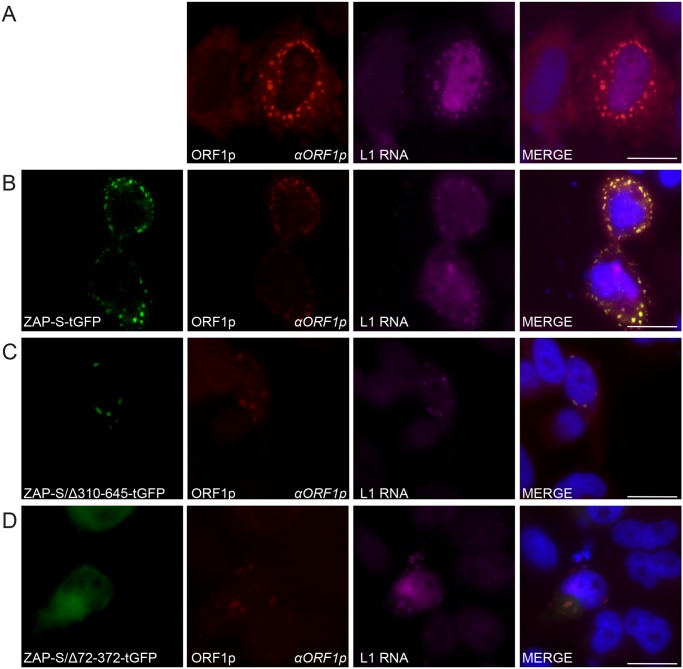
Fig 6. The co-localization of ZAP with L1 RNA and ORF1p in HeLa cells.
(A) Co-localization of transfected L1 RNA and ORF1p: ORF1p (red) expressed from pJM101/L1.3 co-localizes with L1 RNA (magenta). (B) Co-localization of transfected L1 RNA and ORF1p with transfected ZAP-S-tGFP in cytoplasmic foci: ORF1p (red) and L1 RNA (magenta) expressed from pJM101/L1.3 co-localize with ZAP-S-tGFP (green). (C-D) The ZAP-S zinc-finger domain is necessary for co-localization with ORF1p: ORF1p (red) and L1 RNA (magenta) expressed from pJM101/L1.3 co-localize with ZAP-S/Δ310-645-tGFP (green) (panel C). ZAP-S/Δ72-372-tGFP (green) diffusely distributes throughout the cytoplasm, while ORF1p (red) expressed from pJM101/L1.3 forms cytoplasmic foci with L1 RNA (magenta) (panel D). The right-most image of each panel represents a merged image. The name of the protein or RNA is indicated at the bottom left, and the name of the primary antibody used (italicized) is annotated at the bottom right of each image. Nuclei were stained with DAPI (blue) and the scale bar represents 25 μM. Experiments were repeated three times with similar results.

