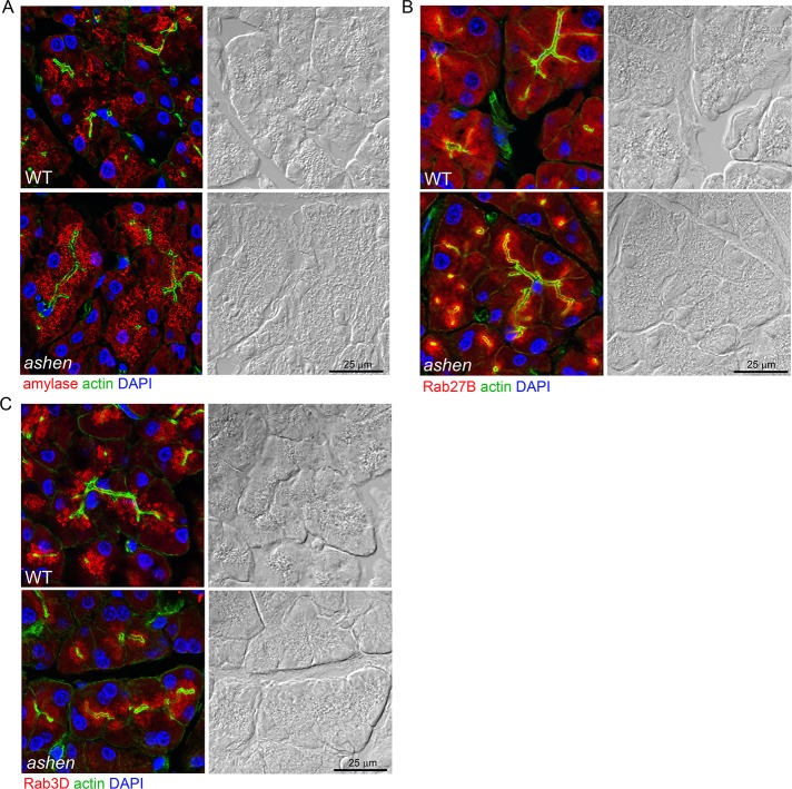
Fig 3. Rab27A deficiency did not affect the localization of zymogen granules, Rab27B and Rab3D.
Pancreas tissues were cryosectioned at 10 μm, stained with phalloidin to label actin (green), DAPI to label nuclei (blue) and anti-amylase (A), anti-Rab27B (B) or anti-Rab3D (C) antibodies (red), respectively. Images were obtained using confocal microscopy. Each panel is paired with the Nomarski image for the same section. Scale bar = 25 μm.

