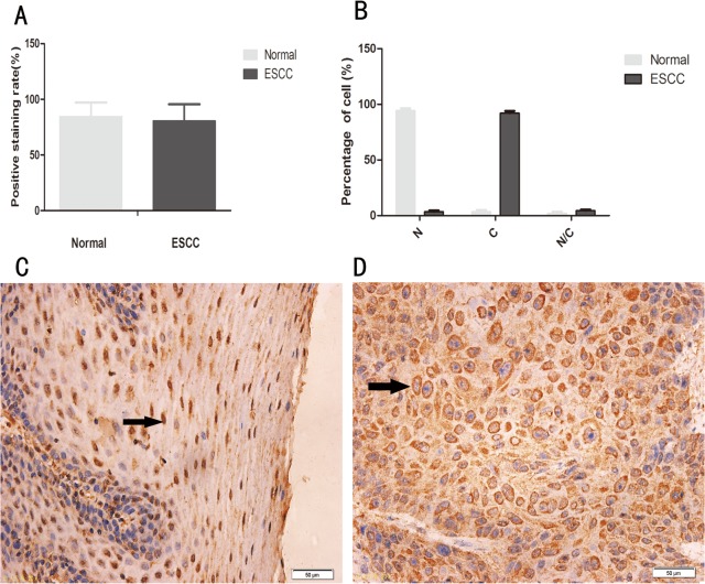
Fig 1. Expression and subcellular localization of ESE3 in normal human esophageal epithelial tissues and ESCC tissues.
(A) Positivity rate in normal human esophageal epithelial tissues and ESCC tissues (P>0.05). (B) Subcellular localization of ESE3 in normal esophageal epithelial cells and ESCC cells. N indicates nuclear localization, C indicates cytoplasmic localization, and N/C indicates nuclear/cytoplasmic localization (equal distribution or uncertain localization). The white bar represents normal esophageal epithelial cells, and the black bar represents ESCC cells (P<0.05). (C) Positive staining of ESE3 in the nucleus of normal esophageal epithelial cells. (D) Positive staining of ESE3 in the cytoplasm of ESCC cells.

