Figure 5.
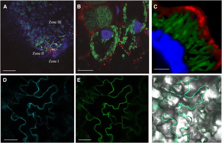
Subcellular localization of MtNramp1-HA. A, Cross section of a 28-dpi M. truncatula nodule infected with S. meliloti constitutively expressing GFP (green) and transformed with a vector expressing the fusion MtNramp1-HA under the regulation of its endogenous promoter. MtNramp1 localization was determined using an Alexa-594-conjugated antibody (red). DNA was stained with DAPI (blue). Infection threads are indicated with asterisks. B, Detailed view of rhizobia-infected cells in zone II. C, Three-dimensional reconstruction of a MtNramp1-HA-expressing cell. GFP-expressing S. meliloti cells are shown in green; red indicates the position of MtNramp1-HA, and blue is DAPI-stained DNA. D, Localization of plasma membrane marker pm-CFP transiently expressed in tobacco leaf cells. E, Localization of MtNramp1-GFP transiently expressed in the same cells. F, Overlay of pm-CFP and MtNramp1-GFP localization in tobacco leaf cells. Bars = 100 µm (A), 15 µm (B), 5 µm (C), and 50 µm (D–F).

