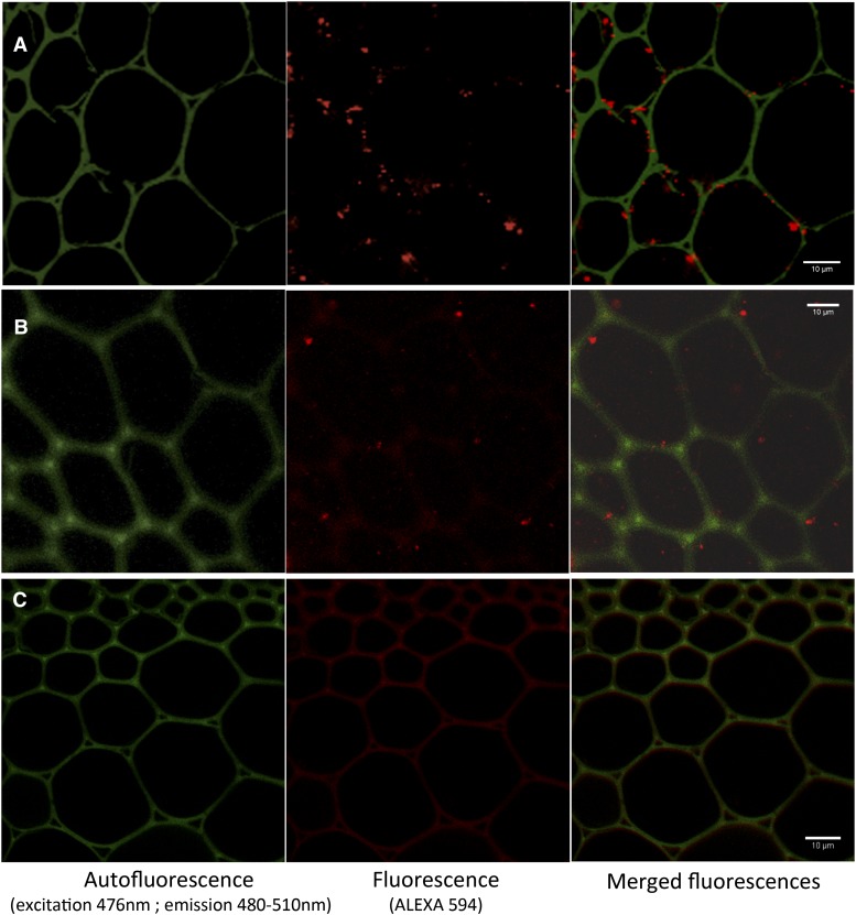
Figure 5.
Subcellular localization of BdLAC5 and BdLAC6 in lignifying interfascicular fibers. Immunolocalizations are imaged with confocal microscopy. A, Immunolocalization of Alexa Fluor 594 after the detection of BdLAC5 primary antibody. B, Immunolocalization of Alexa Fluor 594 after the detection of BdLAC6 primary antibody. C, Control immunofluorescence with Alexa Fluor 594 secondary antibody without primary antibody. Green indicates autofluorescence of cell wall (excitation, 476 nm; emission, 480–510 nm), and red indicates fluorescence of Alexa Fluor 594. Bars = 10 μm.

