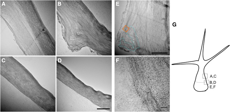
Figure 4.
TEM micrographs of wild-type and exo70H4-1 trichomes. A, Wild-type cell wall above the OR has two distinct layers. B, Wild-type cell wall at the OR. Above the OR, two layers can be recognized, but only one layer is visible beneath the OR. C, exo70H4-1 cell wall at the place corresponding to A. Only one cell wall layer is visible. D, exo70H4-1 cell wall at the place corresponding to B. No OR is visible. E, The blue dotted line highlights the region that forms the OR according to the immunogold labeling. The orange square represents the approximate position of F. F, Detail on the immunogold labeling by the anticallose antibody. The position of F is highlighted by the orange square in E. G, Position and orientation of micrographs depicted for better orientation. Bars = 2 µm (A–E) and 100 nm (F).

