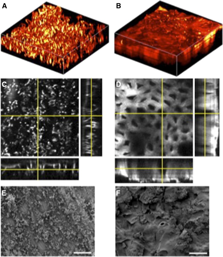
Figure 5.
SRS images and scanning electron micrographs of cuticle of banana and of silver dollar plant. The surface of banana and of silver dollar plant leaves were imaged using SRS microscopy (A–D) and SEM (E and F). A, An SRS image of the adaxial leaf surface of surface banana constructed from an image stack from a 64- × 64-µm field of view. B, An SRS image of silver dollar plant leaf reconstructed from an image stack from a 126- × 126-µm field of view. C and D, Orthogonal views projected from the image stacks presented in A and B, respectively, showing the depth profile of cuticles. E and F, SEM images of the cuticles of banana and silver dollar plant leaves, respectively.

