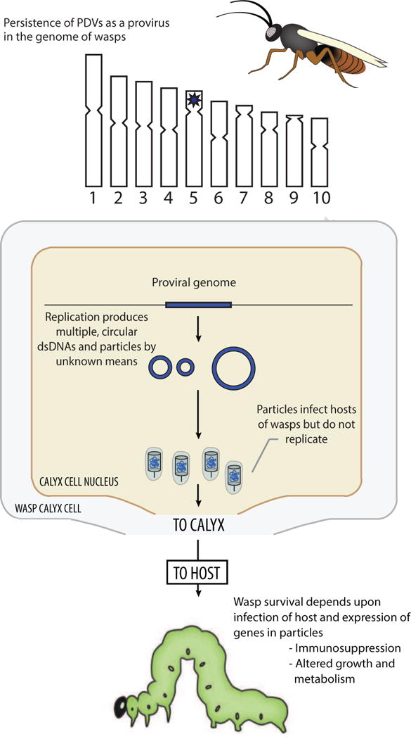
Fig. 1.

Key characteristics of PDVs in 1991 when the family Polydnaviridae was formally recognized by ICTV. The upper part of the figure shows an adult female wasp whose hypothetical genome consists of ten chromosomes. Data at this time indicated that each PDV associated with a given wasp species was genetically distinct and persisted as an integrated provirus. Based on other known dsDNA viruses with a proviral phase, PDV proviral genomes were implicitly assumed to persist as a large, linear dsDNA that was integrated in the wasp genome (*). The middle part of the figure shows the nucleus of a calyx cell. Data showed that particles packaging multiple circular dsDNAs were produced in calyx cells by unknown means followed by storage of particles in a domain referred to as the calyx. The lower part of the figure shows a larval stage lepidopteran host. Data generated prior to 1991 showed that wasps inject PDV particles into hosts, which infect different types of cells and express genes that cause physiological alterations wasp offspring depend upon for survival. Data generated prior to 1991 also showed that no replication of PDVs occurs in the hosts of wasps.
