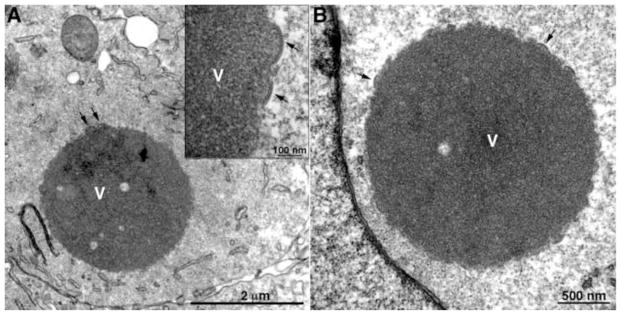
Fig. 3.

Transmission electron microscopic images of a BS-C-1 cell infected with a VACV L2 deletion mutant showing dense inclusions. Arrows point to short crescents. V, dense inclusion of viroplasm. Inset, high magnification of portion of inclusion. Scale bars shown. Adapted from (Maruri-Avidal et al., 2013a)
