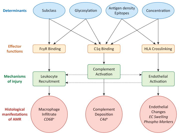
Figure 5. Mechanisms of DSA in graft pathogenesis.
Features of antibody-antigen and antibody-effector system interactions that influence pathogenic functions and mechanisms of injury are shown. Variable factors regulating antibody-antigen interactions (blue ovals) directly influence the capacity of an antibody to trigger effector functions (green boxes), and mechanisms causing graft injury (purple boxes), which ultimately manifest in the graft as common histological features (red bursts). Linear effects are indicated by solid arrows.
The functional endpoints of antibody-mediated injury are interrelated (with potential inflammatory loops indicated by dashed arrows), and likely synergize to cause maximal inflammation during AMR. For example, direct endothelial cell activation by HLA antibodies triggers adhesion of leukocytes, which can be enhanced when those leukocytes bind antibody through FcγRs. Activation of complement at the endothelial cell surface may cause production of anaphylatoxins C3a and C5a, which can act on leukocytes as chemoattractants, or enhance endothelial activation.

