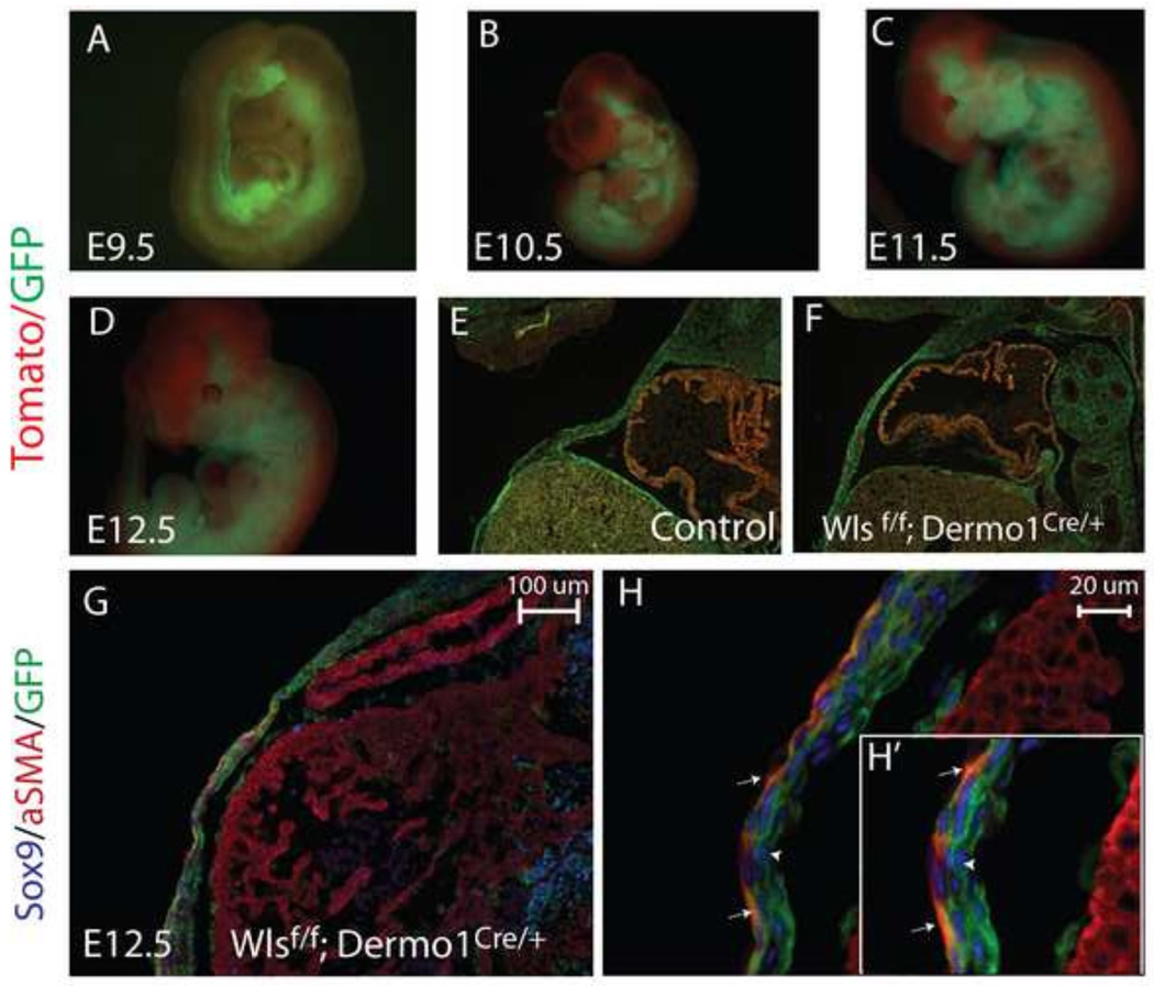
Figure 2. Efficiency and distribution of Dermo1Cre-mediated mesenchymal recombination.
Whole mount images (A–D) and longitudinal sections (E, F) of Wlsf/f;Dermo1Cre/+;Rosa mT/mG embryos are shown. Dermo1Cre-mediated recombination is detected in the mesenchyme as early as E9.5 and progressively expands throughout the mesenchyme as development progresses (A–D). At E12.5, Dermo1Cre-mediated recombination in the body wall is extensive, as demonstrated by the GFP signal in both control and Wlsf/f;Dermo1Cre/+ embryos (E,F). Cre recombination, detected using GFP, was present in both aSMA (white arrows) and Sox9 (white arrowhead) stained cells of the ventral body wall (G,H and H’ insert).

