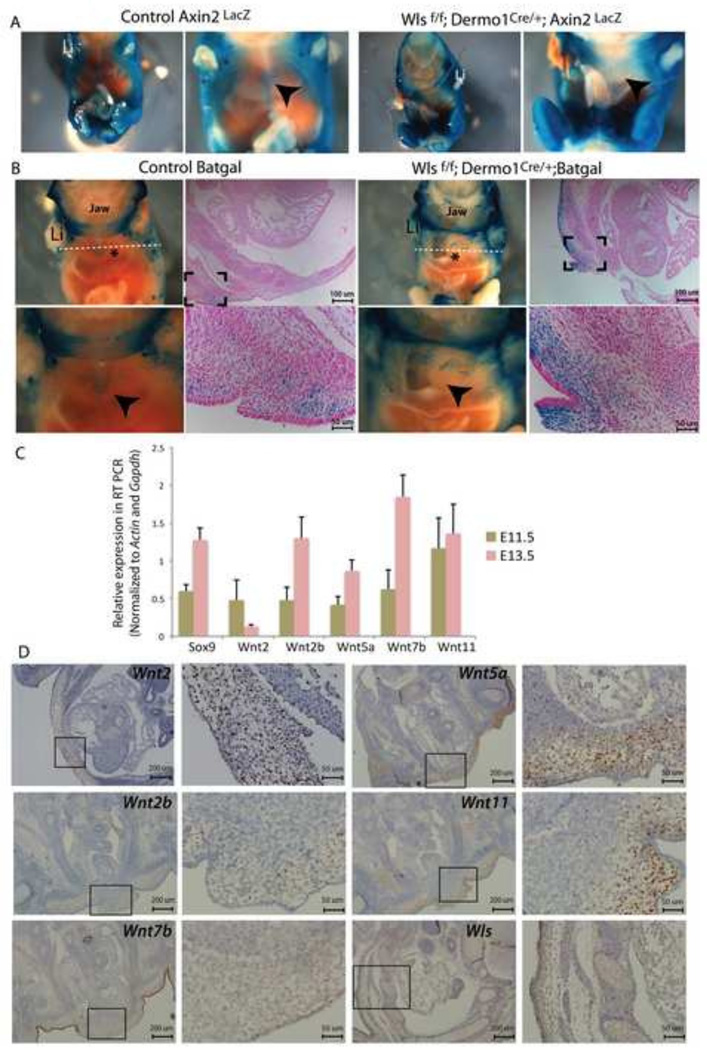
Figure 3. Wnt/β-catenin signaling is operative in ventral body wall.
Canonical Wnt signaling was assessed in E13.5 Wlsf/f;Dermo1Cre/+ embryos. In control embryos, LacZ staining was detected in the ventral body wall converging at the midline as a well-defined stripe (arrowheads in A and B control). In the Wls Wlsf/f;Dermo1Cre/+;Axin2LacZ (A) and Wlsf/f;Dermo1Cre/+;BatGal embryos (B), staining was scattered throughout the ventral body wall, and definitive midline staining was not detected (arrowhead in A and B Wlsf/f;Dermo1Cre/+ panels). Cross sections of the whole mounts show the extent of Wnt/β-catenin signaling in the ventral body wall of control and Wlsf/f;Dermo1Cre/+;BatGal embryos, (B). White dotted lines indicate the plane of section (B). Levels of Wnt ligand mRNA in body wall tissue were determined at E11.5 and E13.5 by RT-PCR (C). Cross sections of E13.5 embryos were hybridized with riboprobes to detect mRNA for Wnt ligands Wnt2, Wnt2a and Wnt7b, Wnt5a and Wnt11, as well as Wls transcripts. Low magnification and high magnification of the areas in squares are shown for each staining. Wls mRNA was detected in both epithelium and mesenchyme, including developing ribs and muscle of the thoracic body wall (D). Li= limb*=heart

