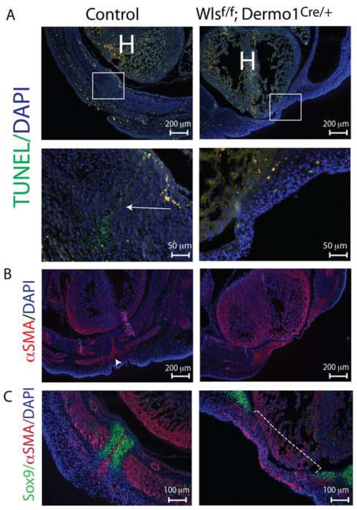
Figure 4. Decreased apoptosis in ventral body wall and impaired migration to midline after mesenchymal loss of Wls.
TUNEL staining was performed on cross and longitudinal sections of E14.5 control and Wlsf/f;Dermo1Cre/+ embryos (A). Note the cell death at the midline (corresponding to the region between the fusing sternal bars) in the control body wall (arrow). This pattern is absent in mutants. Representative images and higher magnifications of areas in square are shown. The midline region were apoptosis takes place in control embryos (A) overlaps with a region that stains positive for αSMA (B). Cross sections of E14.5 embryos stained with Sox9 and αSMA antibodies demonstrate the extent of the lack of fusion of sternal bars in mutants (dotted line) and the position of αSMA stained cells between the sternal bars in control embryos (C). H=heart.

