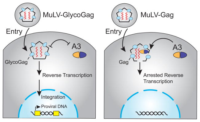
Figure 3.
Model for glyco-Gag protection from restriction by murine A3. The left image depicts glyco-Gag as an oblong blue shape that prevents A3 from accessing reverse transcription complexes. Capsids are primarily composed of the classical Gag (circles not depicted on the left panel). The right panel depicts Gag as a blue oval that causes the capsid to be more loosely formed and susceptible to the A3-mediated block in reverse transcription. Although low levels of G-to-A mutation have been reported, these changes are a minor outcome of A3 activity and are not depicted for clarity. See the text for additional details.

