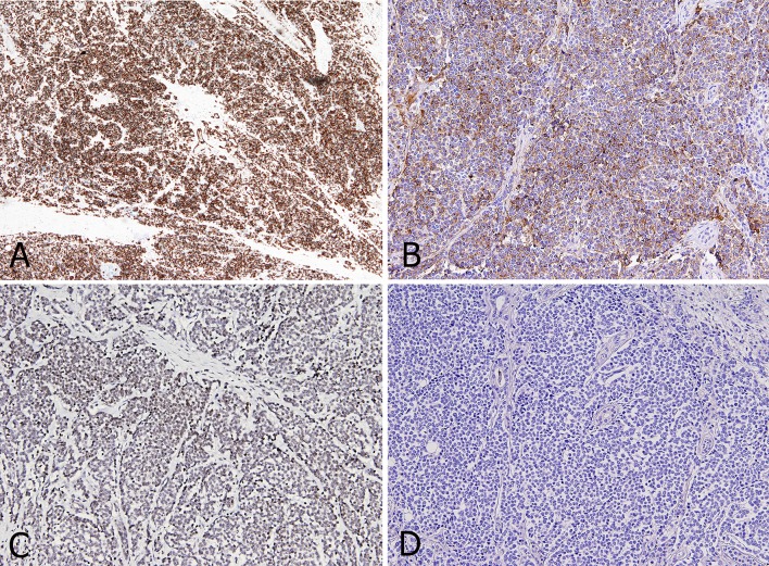
Fig. 3.
Tumor cells show strong and diffuse positivity for CK14 staining (4x) (a) and cytoplasmic and peri-nuclear positivity for Chromogranin A staining (10x) (b). MCPyV staining (10x): granular nuclear positivity for specific Merkel Cell polyomavirus large T antigen (c). CK20 staining (10x): tumor cells are completely negative (d)

