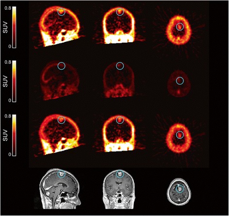
Figure 5.

Image data for patient 10. The image data show radioactivity distribution in normal brain and cerebral metastases (enclosed in blue circle) (top panel) and are separated into non-blood (middle upper panel), blood (middle lower panel) and corresponding contrast-enhanced MRI images (bottom panel). Since the blood volume in metastases may not be 5%, a blood volume fraction model was fitted to dynamic data on a voxel-by-voxel basis. The uptake of radioactivity in the brain metastases was higher than that contributed from a model-fitted blood volume. MRI, magnetic resonance imaging; SUV, standardised uptake value.
