
Fig. 4.
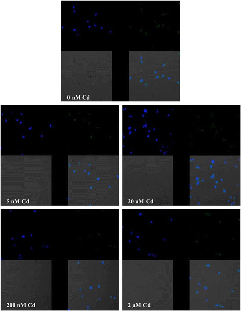
Imaging of COX-2 enzyme by fluorescence microscopy in macrophages cultured with cadmium. Monocytes/macrophages were cultured with Cd solutions for 48 h. The immunohistochemistry was performed using specific primary antibody, mouse anti-COX-2 (the overnight incubation at 4 °C), and secondary antibodies conjugated with flouorochrome–anti-mouse IgG FITC (incubation for 45 min at room temperature). The nuclei of cells were DAPI stained. Image analysis was performed with a fluorescent microscope using filters 38 HE GFP for green fluorescence and 49 DAPI for blue fluorescence