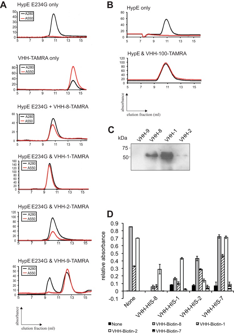
FIGURE 2.
Validation and characterization of HypE-specific VHHs. Recombinant HypE(187–437) E234G (A) and HypE(187–437) (B) were incubated with TAMRA-labeled VHHs. Complex formation was assessed by size exclusion chromatography. Black lines (A280) represent total protein, and red lines (A545) represent the TAMRA signal. C, endogenous HypE was immunoprecipitated from HeLa cell lysates using VHH-coupled beads and analyzed by Western blotting with a commercial anti-HypE antibody. D, competition ELISAs were performed using His6-VHHs for initial epitope masking and biotin-VHH for subsequent probing.

