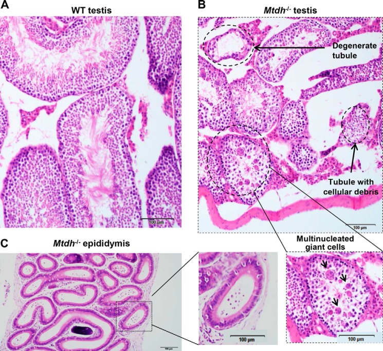
FIGURE 2.
Testes from homozygous Mtdh exon 3-deficient male mice display testicular degeneration and a lack of mature spermatozoa. A and B, morphology of testes from WT mice (A) and homozygous Mtdh exon 3-deficient mice (B) by H&E staining. Multinucleated giant cells are indicated by arrows. C, H&E staining of a representative section of the epididymis from an Mtdh exon 3-deficient mouse.

