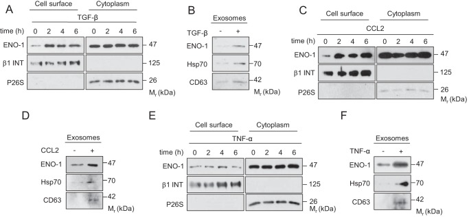
FIGURE 3.
ENO-1 exteriorization is triggered by stimuli promoting tumor progression. A, C, and E, cell surface expression of ENO-1 in MDA-MB-231 cells exposed to 20 ng/ml TGF-β1 (A), 100 ng/ml CCL2 (C), or 50 ng/ml TNF-α (E) for the indicated time points. The purity of cytosolic and cell membrane fractions was assessed by probing the samples for β1 INT and P26S, respectively (n = 3). Representative Western blots are shown. B, D, and F, abundance of ENO-1 in exosomes isolated from MDA-MB-231 cells exposed to 20 ng/ml TGF-β1 (B), 100 ng/ml CCL2 (D), or 50 ng/ml TNF-α (F) as assessed by Western blotting. Hsp70 and CD63 served as exosome markers (n = 3). Representative Western blots are demonstrated.

