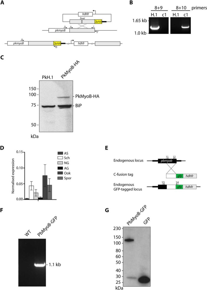
FIGURE 2.
Generation of PkMyoB-HA and PbMyoB-GFP and detection of myob throughout the P. berghei life cycle. A, diagram representing C-terminal tagging of Pkmyob with a triple HA tag. Primers shown (arrows) were used to amplify the region of homology (6 and 7) for diagnostic PCR of WT parasite sequences (8 and 9) or parasites where integration had taken place (8 and 10). B, diagnostic PCR on genomic DNA showing integration of PkMyoB-HA into the myob locus in clone c1 with parental H.1 DNA as a control. C, Western blot with an anti-HA antibody on parental PkH.1 and PkMyoB-HA parasite lysates. In PkMyoB-HA parasites, a protein of ∼90 kDa is detected. The same blot was probed with an antibody against BiP to demonstrate equivalent sample loading. D, quantitative RT-PCR to show mRNA expression of Pbmyob during the parasite life cycle. Arginyl-tRNA synthetase and hsp70 were used as endogenous controls for normalization. Bars, three biological replicates, each ± S.E. AS, all asexual blood stages; Sch, schizonts; NG, nonactivated gametocytes; AG, activated gametocytes; Ook, ookinetes; Spz, sporozoites. E, diagram representing C-terminal tagging of PbMyoB with GFP by single homologous recombination into endogenous myob gene, showing primers 11 and 12 to amplify the region of homology, and primers 13 and 14 to detect integration. F, confirmation of integration by PCR of Pbmyob-gfp using primer pair 13 and 14. G, Western blot of extracts of parasites expressing PbMyoB-GFP or GFP.

