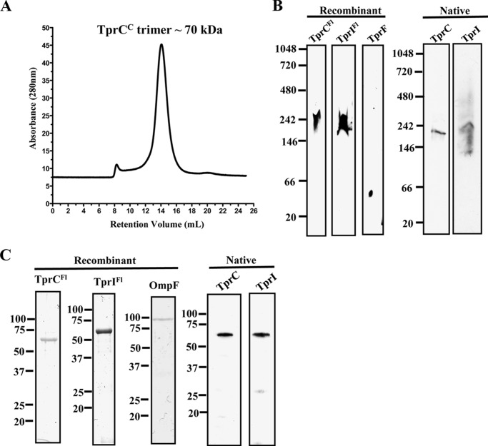
FIGURE 5.
TprCFl and TprIFl form SDS-dissociable trimers. A, size exclusion chromatography analysis of TprCC in 50 mm Tris, pH 8, 200 mm NaCl, and 0.1% lauryldimethylamine oxide. The relative size of the TprCC was estimated by comparing the retention volume with a standard curve calculated using blue dextran. B, blue native PAGE. TprCFl, TprIFl, TprF, and T. pallidum lysates solubilized in 0.5% DDM with 50 mm Tris (pH 7.0) were separated by blue native PAGE followed by immunoblot analysis with antisera directed against TprCFl (TprCFl and TprIFl), TprCN (TprF), TprCSp (native TprC), and TprISp (native TprI). C, SDS-PAGE without boiling. TprCFl, TprIFl, OmpF, and T. pallidum lysates were subjected to SDS-PAGE without boiling. Recombinant proteins were stained with GelCode®, whereas T. pallidum lysates were immunoblotted with TprCSp and TprISp antisera. Molecular mass standards (kDa) are shown on the left.

