FIGURE 3.
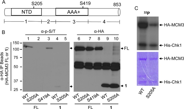
Identification of MCM3 phosphorylation sites. A, schematic diagram of MCM3 FL and four fragments (1–4). AAA+, ATPase domain; NTD, nucleic acid-binding domain. B, HEK293T cells were transfected with HA-MCM3 FL (WT, S205A, or S419A) or fragment 1 (WT or S205A) for 48 h, IPed with anti-HA antibodies, and blotted with the anti-Ser(P)/Thr(P) mixture (left). The same membrane was stripped and reblotted with anti-HA antibodies (right). C, HA-MCM3 WT or S205A mutant was collected from transfected HEK293T cells, treated with CIAP, and used as the substrate for in vitro kinase assay in the presence of [γ-32P]ATP. The SDS-polyacrylamide gel was stained with Coomassie Blue (lower), dried, and exposed to x-ray radiography (upper).

