FIGURE 5.
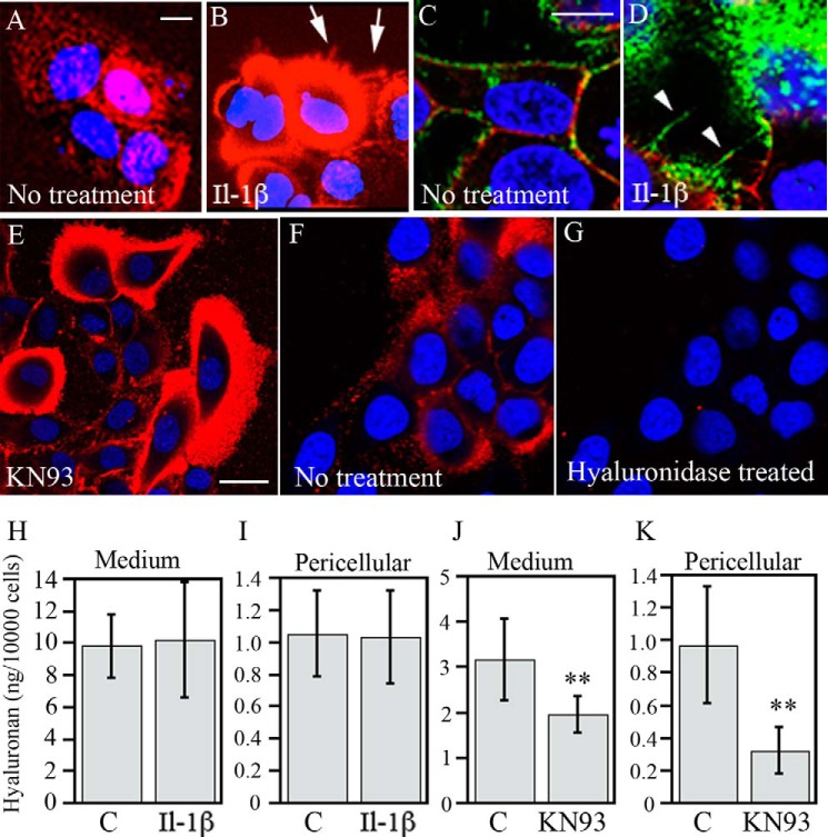
IL-1β and inhibition of CAMKII induce the formation of extended hyaluronan-coats. HaCaT cells were treated with either IL-1β or KN93 for 20 h. A, B, E, F, and G, staining of live cultures for hyaluronan in situ with 5 μg/ml Alexa Fluor 568-labeled HABC in the culture medium. C and D, fixed cultures stained with biotinylated HABC and the anti-CD44 antibody Hermes 3, visualized by FITC-streptavidin and TR-labeled secondary antibody, respectively. Red in A, B, E, F, and G and green in C and D, hyaluronan. Red in C and D, CD44; blue (DRAQ5TM dye), nuclei. Cell protrusions and hyaluronan cables are indicated by arrows and arrowheads, respectively. Scale bars, 10 μm (A–D) and 20 μm (E). F and G, specificity of HABC label was checked with hyaluronidase treatment prior to staining. H and J, hyaluronan content in the culture medium. I and K, pericellular hyaluronan (released by trypsin). The treatment time was 20 h for IL-1β and 6 h for KN-93. The values represent means ± S.E. (error bars) from five (IL-1β) and three (KN93) independent experiments. **, p < 0.01 (ANOVA).

