Figure 4.
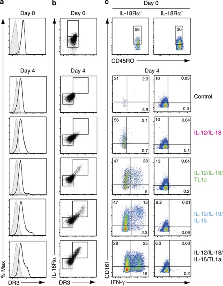
The tumor necrosis factor-like cytokine 1A (TL1a) receptor death receptor 3 (DR3) is expressed at high levels on interleukin-18 receptor alpha (IL-18Rα)+CD45RO+CD4+ T cells in cytokine cultures. (a–c) DR3, IL-18Rα, CD161, and interferon-γ (IFN-γ) expression was assessed on peripheral blood CD45RO+CD4+ T cells by flow cytometry at day 0 or day 4 after culture (1 × 10⁶ cells ml−1, 200 μl per well) with interleukin-12 (IL-12) (2 ng ml−1), IL-18 (50 ng ml−1), IL-15 (25 ng ml−1), and TL1a (100 ng ml−1) or medium alone (control). (a) Representative plots showing DR3 expression (black line, unfilled histogram) or fluorescence minus one (FMO) control (shaded histogram) on CD45RO+CD4+ T cells. (b) DR3 and IL-18Rα expression on CD45RO+CD4+ T cells. Boxes within plots are gates defining IL-18Rα+ or IL-18Rα− (day 0) and IL-18Rα+DR3hi or IL-18Rα−DR3lo (day 4) CD4+ T cells. (c) IFN-γ and CD161 expression on IL-18Rα+DR3hi or IL-18Rα−DR3lo CD4+ T cells as defined in b. Quadrants are set based on isotype control (IFN-γ) and FMO control (CD161) staining of IL-18Rα+DR3hi and IL-18Rα−DR3lo CD4+ T cells under each of the cytokine culture conditions. Results are representative of 3 (day 0) or 7 (day 4) biological replicates.

