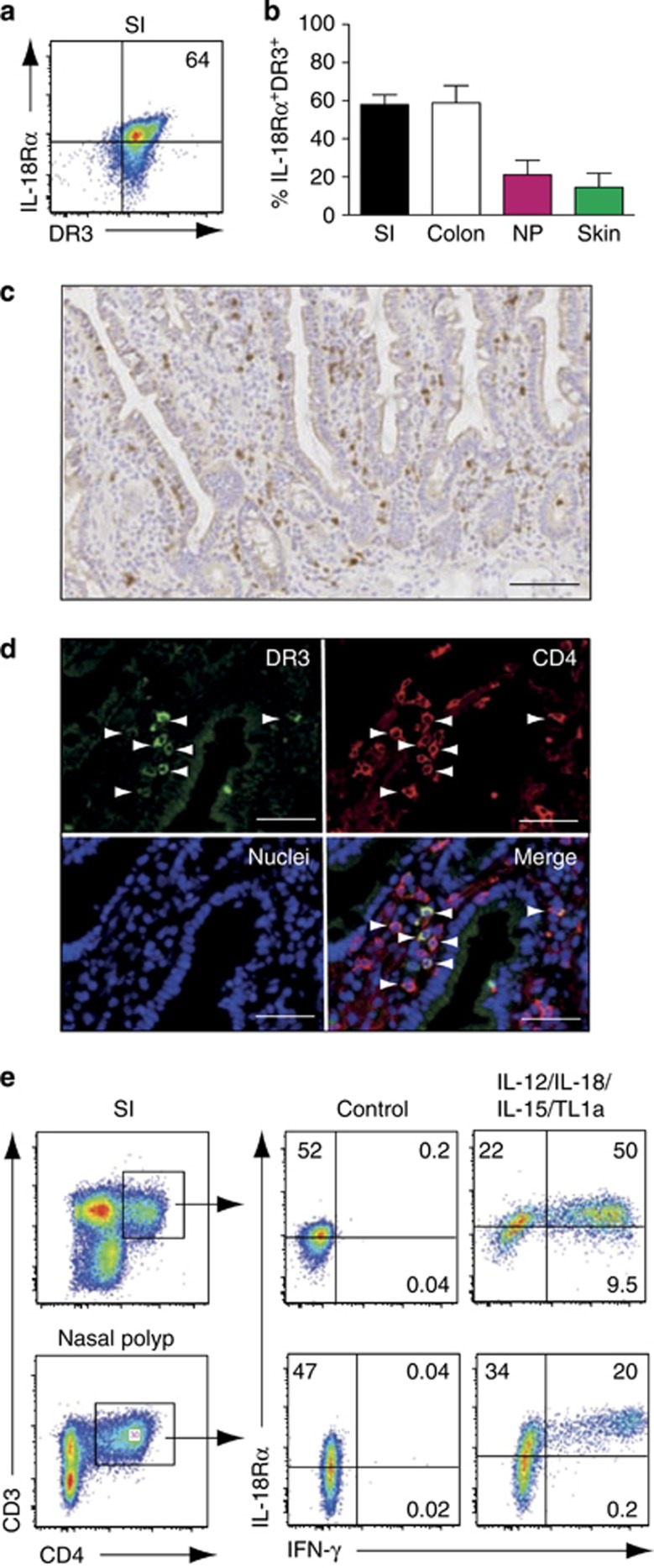
Figure 6.
Interleukin-18 receptor alpha (IL-18Rα)+death receptor 3 (DR3)+CD4+ T cells are present at barrier surfaces. (a) Representative flow cytometry plot and (b) percentage of IL-18Rα+DR3+ cells in the indicated organs. IL-18Rα and DR3 expression on CD4+ T cells was assessed after incubation in culture medium for 2 days to allow re-expression of DR3. Gating is on live, CD3+CD4+ cells. (b) Results are mean (s.e.m.) of 7 (small intestine, SI), 3 (nasal polyp, NP), 5 (colon), and 3 (skin) stainings performed. (c and d) DR3+CD4+ cells are diffusely distributed throughout the healthy human small intestine. (c) Small intestinal sections were stained with DR3 antibody. (d) Sections were stained with DR3 and CD4 antibody together with 4',6-diamidino-2-phenylindole (DAPI) to identify cell nuclei and analyzed by confocal microscopy. Arrowheads depict DR3+CD4+ cells. Bars=(c) 0.1 mm and (d) 50 μm. Images are from one representative tissue of at least (c) 20 and (d) 3 analyzed. (e) Cytokines induce tissue-resident IL-18Rα+CD4+ T cells to produce interferon-γ (IFN-γ). SI and NP cell suspensions were cultured in medium alone (control) or with interleukin (IL)-12 (2 ng ml−1), IL-18 (50 ng ml−1), IL-15 (25 ng ml−1) and tumor necrosis factor-like cytokine 1A (TL1a) (100 ng ml−1) for 2 days before analysis. Cells were gated on live CD3+CD4+ cells (left panels). Results are representative of 5 (small intestine) and 3 (NP) biologic replicates.

