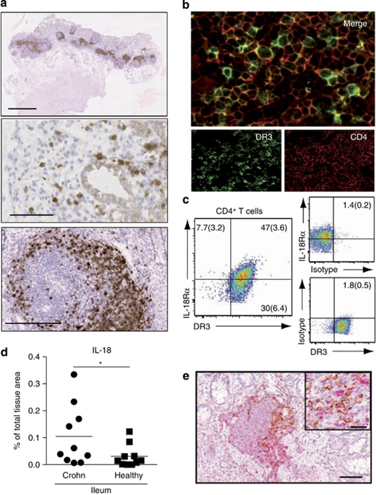
Figure 8.
Interleukin-Il-18R alpha (IL-18Rα)+death receptor 3 (DR3)+CD4+ T cells colocalize with interleukin-18 (IL-18)-expressing cells in inflamed small intestine. (a) DR3+ cells (brown) are diffusely distributed throughout the inflamed human small intestine and predominantly located within lymphoid aggregates and follicles. Six micrometer sections of inflamed small intestine biopsies from Crohn's disease (CD) patients were stained with DR3 antibody as described in Methods section. Results are from one representative patient of 15 analyzed. Bars=2 mm (top), 0.1 mm (middle), and 0.25 mm (bottom). (b) DR3+ cells within aggregates are CD4+ T cells. Sections were stained with DR3 and CD4 antibody as described in Methods section and analyzed by confocal microscopy. Results are from one representative patient of 5 analyzed. (c) Most DR3+CD4+ T cells from inflamed small intestine coexpress IL-18Rα. Lamina propria mononuclear cells were prepared from inflamed small intestine and maintained in culture medium for 2 days before analysis. Gating is on live CD3+CD4+ cells. Results are mean (s.e.m.) of five biological replicates (two from surgical resection tissue and three from biopsies of untreated, newly diagnosed CD patients). (d) IL-18 expression increases in the inflamed small intestine of CD patients. Tissue sections were stained by immunohistochemistry for IL-18 and the numbers of pixels displaying IL-18-positive staining were quantified using computerized image analysis, as described in Methods section. Data are expressed as mean percentage of the total tissue area occupied by IL-18-positive immunoreactivity (circles denote individual patients) (e) IL-18-producing cells colocalize with CD4+ cells in lymphoid aggregates in CD small intestine. Sections were stained with CD4 antibody (brown) and IL-18 antibody (red). Results are a representative staining from 1 of 10 patients examined. Bar=0.1 mm. Insert bar=40 μm.

