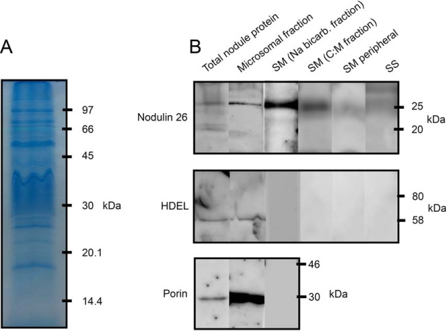
Fig. 1.
One dimensional SDS-PAGE of SM proteins and Western blot analysis of nodulin 26, HDEL and porin in nodule fractions. A, Ten micrograms of sodium bicarbonate stripped symbiosome membrane (SM) protein resolved on a 12% SDS-polyacrylamide gel and stained with Coomassie brilliant blue. B, Ten micrograms of protein from total nodule, microsomal, sodium bicarbonate stripped SM, C:M extracted SM, SM peripheral, and peribacteroid space (PBS) fractions were resolved on 12% SDS-polyacrylamide gels then transferred to PVDF membranes. Blots were blocked and probed with antibodies for either nodulin-26, HDEL or porin proteins.

